Hussam AS Murad1,2 ![]() ,
Hamid SA Habib3,
Yasser M Kamel4.5,
Salah A Alsayed6,
Soad S Ali7,
Zohair G Gazzaz8
,
Hamid SA Habib3,
Yasser M Kamel4.5,
Salah A Alsayed6,
Soad S Ali7,
Zohair G Gazzaz8
For correspondence:- Hussam Murad Email: muradha2000@yahoo.com Tel:+966541541341
Received: 6 May 2016 Accepted: 21 August 2016 Published: 30 September 2016
Citation: Murad HA, Habib HS, Kamel YM, Alsayed SA, Ali SS, Gazzaz ZG. Oral thearubigins do not protect against acetaminophen-induced hepatotoxicity in mice. Trop J Pharm Res 2016; 15(9):1909-1914 doi: 10.4314/tjpr.v15i9.14
© 2016 The authors.
This is an Open Access article that uses a funding model which does not charge readers or their institutions for access and distributed under the terms of the Creative Commons Attribution License (http://creativecommons.org/licenses/by/4.0) and the Budapest Open Access Initiative (http://www.budapestopenaccessinitiative.org/read), which permit unrestricted use, distribution, and reproduction in any medium, provided the original work is properly credited..
Purpose: To investigate the potential protective effect of oral repeated doses of thearubigins against acetaminophen-induced hepatotoxicity in mice.
Methods: Mice were randomly divided into six groups (n=8) and administered the following: Control group (saline), acetaminophen group (saline), N-acetylcysteine group (500 mg/kg/day), and thearubigins groups (60, 70, 100 mg/kg/day). The drugs were given orally by gavage for seven days. On day 7, 1 h after the last dose of treatment, the mice (except control group) were given a single dose of acetaminophen (n-acetyl-p-aminophenol, APAP) orally by gavage (350 mg/kg) and then sacrificed 4 h post-APAP intake. Blood was collected for biochemical measurements and their liver were subjected to biochemical and histopathological assessment.
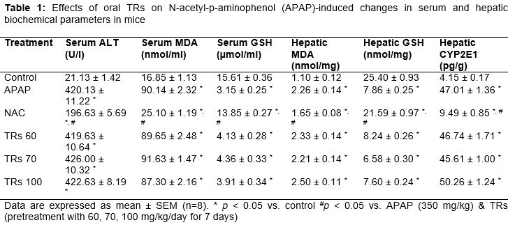
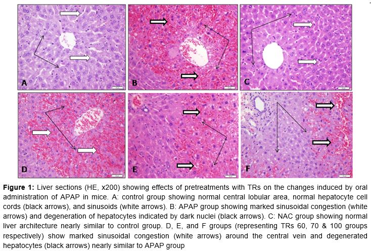
Results: The acetaminophen group showed significant increases (p < 0.001) in serum alanine aminotransferase level, hepatic cytochrome P2E1 level, and serum and hepatic malondialdehyde levels. Moreover it showed significant decrease (p < 0.001) in serum and hepatic glutathione levels. Morphologically, the liver sections showed cellular necrosis, vacuolization, and degeneration around the centrilobular veins. Pretreatment with N-acetylcysteine reversed all acetaminophen-induced changes (p < 0.001 for all biomarkers except for hepatic MDA (p = 0.014) while pretreatment with thearubigins failed to reverse any of them.
Conclusion: Oral repeated doses of thearubigins failed to protect against acetaminophen-induced hepatotoxicity in mice and didn't affect hepatic cytochrome P2E1 level.
Introduction
Acetaminophen (APAP) is safe when used in regular doses where nearly 95 % of it is metabolized in the liver by glucuronidation and sulfation to non-toxic metabolites. Only 5 % is oxidized by CYP450 isozymes (mainly CYP2E1) to n-acetyl-p-benzoquinone imine (NAPQI) which is then reduced by glutathione (GSH) and excreted as mercapturic acid [1]. In overdose, acetaminophen causes acute liver failure due to saturation of the sulfation pathway and shifting of more APAP to the CYP450 pathway forming excessive NAPQI. This excessively-produced NAPQI severely depletes cellular GSH and covalently binds to cellular proteins with increased formation of free radicals causing mitochondrial damage, cellular necrosis, and organ failure. In addition, the increased production of free radicals causes mucosal injuries, damage of cell membrane, and cell death by attacking cellular lipid constituents leading to activation of lipid peroxidation and increasing malondialdehyde (MDA) levels. Antioxidants and GSH precursors such as N-acetylcysteine (NAC) plays an important role in the treatment of APAP toxicity through antagonizing oxidative stress and promoting tissue regeneration [2-4].
In our recently-published study [5], we found that intraperitoneal single dose thearubigins (TRs); the main phenolic pigment of black tea extract; dose-dependently protected against APAP-induced hepatotoxicity in mice. The effects of toxins and drugs differ according to their route of administration. For example, yessotoxin administered to mice orally was found less toxic than that given by the intraperitoneal route [6]. Also, oral quercetin failed to enhance the antitumor effect of trichostatin A in mice while the intraperitoneally-injected quercetin did [7].
The present study was designed to investigate the potential hepatoprotective effect of oral pretreatment with TRs given to mice for seven days prior to oral administration of APAP on the seventh day.
Methods
Preparation of thearubigins (TRs)
The Black tea extract (BTE) was prepared, its ingredients were determined using high performance liquid chromatography, and aqueous solutions of TRs (60, 70, and 100 mg/kg) were prepared as previously described [8,9]. Briefly, the lyophilized BTE was suspended in water and extracted continuously with chloroform to remove caffeine. The aqueous layer was extracted continuously with ethyl acetate (2 × 40 mL) for removing of theaflavins (TFs). Finally, TRs were extracted with n-butanol (3 × 40 mL) and evaporated using rotary vacuum evaporator.
Animals and drugs
Swiss male albino mice (20 - 30 g) were obtained and housed in cages at 20 – 22 °C room temperature in 12 h light-dark cycle. Food and water were available ad libitum. All drugs and chemicals were purchased from Sigma-Aldrich Corp. (St. Louis, MO, USA) unless stated otherwise.
Induction of acetaminophen toxicity and treatment groups
After acclimatization for a week, mice were randomly divided into six groups (n=8) and administered the following: Control group (saline), APAP group (saline), NAC group (500 mg/kg/day) [10], and three TRs groups (60, 70, 100 mg/kg/day) [11]. Drugs were given orally by gavage for seven days. On the 7th day, 1 h after the last doses of treatments, all groups (except the control group) were given a single dose of APAP by oral gavage (350 mg/kg) [10] and then the mice were sacrificed 4 h post-APAP injection by cervical dislocation under light anesthesia. Blood was collected for biochemical measurements and livers were removed, washed in saline, and used for biochemical measurements and histopathological examination [12].
The protocol of the study was approved by King Abdulaziz University Research Ethics Committee (ref no. 142-15) and adhered to the International Guidelines for the Care and Use of Laboratory Animals [13].
Serum biomarker measurements
The measurements of serum analytes were done by using commercially available kits for alanine aminotransferase (ALT) (Teco Diagnostics, CA, USA), and malonaldehyde (MDA) and GSH (Cell Biolabs, San Diego, CA, USA) according to the manufacturer’s protocol.
Hepatic malonaldehyde (MDA) measurement
The hepatic MDA was measured by using the OxiSelect™ TBARS Assay Kit (Cell Biolabs, Inc., CA, USA). Briefly, liver specimens were homogenized in 1.15 % potassium chloride buffer (1/10 w/v) and centrifuged. The 10 % homogenate was mixed with an aqueous solution of thiobarbituric acid (TBA). After heating at 95 °C for 1 h, the produced red pigment was extracted with butanol and absorbance was measured at 532 nm using a plate reader [14].
Hepatic glutathione (GSH) measurement
The hepatic GSH was measured using GSH assay kit (Cayman Chemical Company, MI, USA). Briefly, liver specimens were homogenized in 5 % trichloroacetic acid and then centrifuged for 30 min at 4 °C. DTNB (5,5'-dithiobis 2-nitrobenzoic acid) was added to the supernatant. The assay involved reduction of DTNB to a yellow product by the sulfhydryl (SH) group of the GSH present in the sample. The absorbance was measured at 405 nm using a plate reader. GSH in the supernatant were quantified using the extinction coefficient of DTNB and normalized to the protein in each sample [15].
Hepatic CYP2E1 measurement
Hepatic CYP2E1 was measured using CYP2E1 assay ELISA kit (Cusabio, Wuhan, China). Briefly, 100 mg liver tissue was rinsed in 1X phosphate buffered saline (PBS), homogenized in 1 mL of 1X PBS, and stored at - 20 °C. Following two cycles of freezing and thawing (to damage the cell membranes) the homogenate was centrifuged for 5 min at 5000 at 4 °C. The supernatant was removed and the CYP2E1 level was measured immediately. Briefly, a complex was formed between the CYP2E1 present in the sample and its specific antibody pre-coated on the wells. A biotin-conjugated antibody specific for CYP2E1 was then added followed by addition of the avidin conjugated horseradish peroxidase (HRP). On adding the substrate solution, a color was developed which was measured using a microplate reader at 450 nm [16].
Histopathological examination
Liver samples were fixed in 10 % phosphate-buffered formalin, embedded in paraffin, and sectioned at 3-5 µm thickness. Liver sections were stained with hematoxylin and eosin (HE), examined in a light microscope (Olympus, BH-2, Tokyo, Japan), and the lesions were graded as mild, moderate, and severe [17].
Statistical analysis
Data are given as mean ± standard error of mean (SEM). Comparison between two groups was made using Student's t-test. One-way analysis of variance (ANOVA) with Tukey’s post-hoc test was applied for comparison of variance between groups. SPSS version 18.0 software was used in the statistical analysis and p < 0.05 was considered statistically significant.
Results
APAP mice showed significant increases in serum ALT, hepatic CYP2E1, and serum and hepatic MDA levels. Moreover, they showed significant decreases in serum and hepatic GSH levels. Morphologically, the liver sections showed cellular necrosis, vacuolization, and degeneration around the centrilobular veins. Pretreatment with NAC (500 mg/kg/day by oral gavage for 7 days) reversed the biochemical and histopathological changes induced by oral administration of APAP (350 mg/kg) on the seventh day while pretreatment with TRs (60, 70, 100 mg/kg/day) in the same schedule failed to reverse these changes ( and ).
Discussion
It is well established that acetaminophen (APAP) overdose in mice causes hepatotoxicity manifested by increases in serum ALT level, hepatic CYP2E1 level, and serum and hepatic MDA levels in addition to decreases in serum and hepatic GSH levels [1,18]. Morphologically, the liver sections showed cellular necrosis, vacuolization, and degeneration around the centrilobular veins.
NAC protects against APAP overdose in mice and reverses the APAP-induced biochemical and histopathological changes because it replenishes hepatic GSH which scavenges NAPQI, helps rapid recovery of mitochondrial GSH, and scavenges reactive oxygen and nitrogen radicals [19].
In the current study, pretreatment of mice with TRs orally by gavage for 7 days failed to reverse the biochemical and histopathological changes induced by oral administration of APAP in the seventh day. These results contrast with our recently-published findings [5] where posttreatment with TRs by intraperitoneal injection dose-dependently reversed the changes induced by prior intraperitoneal injection of APAP. This discrepancy may be attributed to differences in study design, routes of administration, and in time points at which the outcomes were measured (in the current work mice were sacrificed at 4 h post-APAP intake while in the previous work mice were sacrificed at 6 h post-APAP injection).
The mechanisms of APAP-induced hepatotoxicity are complicated and thus on testing a drug for a potentially-protective effect, misinterpretation of the results may occur especially with plant extracts [20]. Perhaps this applies to our studies; the term TRs was introduced since fifty years ago however their exact chemical structure remains undefined up till now [21]. Consequently, no clear data about their pharmacokinetic profiles are available. In addition, there is little research on black tea theaflavins and thearubigins regarding their gastrointestinal absorption, metabolism, and potential impact on health [22]. A similar contrast was reported by Catterall et al [23,24]. In the earlier study [23], they found that theafulvins, a fraction of black tea TRs, inhibited the hepatic CYP450 activity associated with the CYP2E subfamilies. However, in the second study [24], they reported that dietary intake of theafulvins for four weeks failed to affect the hepatic CYP450 activity or composition in rats, but downregulated expression and activity of the intestinal CYP 450 enzymes including CYP2E1. They attributed these contradictory results to poor oral bioavailability of the black tea TRs due to poor absorption and/or extensive first pass intestinal elimination by mammalian and microbial enzymes resulting in higher local concentrations of theafulvins in GIT. A green tea polyphenol given daily for four weeks on an empty stomach; to improve its oral bioavailability; failed to affect the human CYP-mediated drug metabolism at any clinically relevant level [25].
In contrast, tea polyphenols (TPs) given for six days prior to paracetamol (1000 mg/kg) dose-dependently suppressed CYP450 expression, reduced CYP2E1 and CYP1A2 expression at both protein and mRNA levels, relieved paracetamol-induced hepatic injury, and decreased number of deaths in mice with APAP overdose [26]. Moreover, black tea and its main polyphenols effectively scavenged the free radicals in vitro in mice and thus they increased the superoxide dismutase and glutathione peroxidase activities and decreased the MDA level [27]. Selective inhibitors of CYP2E1 such as 4-methylpyrazole and disulfiram significantly reduced NAPQI production and then they could be useful in treating acetaminophen overdose [16]. Chlorogenic acid (a polyphenolic compound abundant in fruits, vegetables, and some medicinal herbs) relieved acetaminophen-induced liver damage through modest inhibition of CYP2E1 and antioxidant effects [12]. The complex structure of TRs is considered an obstacle facing studies that evaluate their bioavailability and mechanisms of action [28], however such studies are necessary to help explain the different; even contradictory; results obtained with changing their route of administration.
Conclusion
Oral repeated doses of black tea TRs do not protect against acetaminophen-induced hepatotoxicity in mice and don't affect hepatic CYP2E1 activity. These findings contradict our recent findings that indicate dose-dependent protective effect of a single intraperitoneal injection of TRs against acetaminophen-induced hepatotoxicity in mice through their antioxidant effect. This discrepancy may be due to poor GI absorption of TRs, thus a full pharmacokinetic study is recommended.
Declarations
Acknowledgement
References
Archives


News Updates